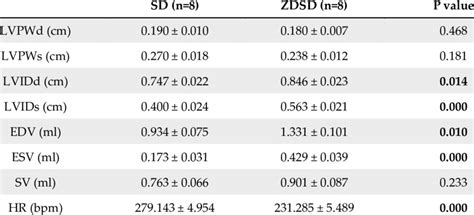
normal lv thickness | left ventricular hypertrophy symptoms normal lv thickness PK G´«Xoa«, mimetypeapplication/epub+zipPK G´«X EPUB/css/aha-circimaging.css•’ÝjÃ0 . $41.99
0 · left ventricular wall thickness chart
1 · left ventricular hypertrophy symptoms
2 · left ventricular hypertrophy risk
3 · left ventricular hypertrophy causes
4 · left ventricle increased wall thickness
5 · heart wall thickness chart
6 · Lv wall thickness normal values
7 · Lv thickness echo
by Vogue Arabia. Presented by chanel. Actress Whitney Peak is the fresh new face of Coco Mademoiselle. Her connection with the maison shows there is more than meets the eye. Les 4 Rouges Yeux Et Joues – Eyeshadow And Blush Palette – 958 Caractere, Rouge Allure – 194 Sensibilite, Chanel. Photo: Baard lunde.
left ventricular wall thickness chart
Increased left ventricular myocardial thickness (LVMT) is a feature of several cardiac diseases. The purpose of this study was to establish standard reference values of normal LVMT with cardiac magnetic resonance and to assess variation with image acquisition plane, .PK G´«Xoa«, mimetypeapplication/epub+zipPK G´«X EPUB/css/aha-circimaging.css•’ÝjÃ0 .

lv pur131 rf filter
Each echocardiogram includes an evaluation of the LV dimensions, wall thicknesses and function. Good measurements are essential and may have implications for therapy. The LV dimensions must be measured . Normal 2D measurements: LV minor axis ≤ 2.8 cm/m 2, LV end-diastolic volume ≤ 82 ml/m 2, maximal LA antero-posterior diameter ≤ 2.8 cm/m 2, maximal LA volume ≤ 36 ml/m 2 (2;33;35). ∗∗ In the absence of other .A normal ejection fraction is 53-73% (52-72% for men, 54-74% for women). Refer to Table 2 (normal values for non-contrast images) and Table 4 (recommendations for the normal range, .
Normal values of the thickness of the compact LV myocardium have been shown to vary by type of pulse sequence (FGRE versus bSSFP) [53, 54]. For the purposes of this .Left and right ventricle. Visual assessment of systolic function. Visual assessment of regional wall motion (left ventricle) Recommended by American Society for Echocardiography (J Am Soc Echocardiogr 18:1440-1463, 2005). Left .
Normal sex- and age-specific reference ranges for left ventricular mid-diastolic wall thickness (LV-MDWT) at prospective electrocardiographically triggered mid-diastolic CT angiography were . Normal values of LV mass were derived from 1,854 subjects. •. The upper limits of normal were established for linear, 2D, and 3D techniques. •. Normal values differ by age, sex, race, and technique used. Abstract. .This document provides updated normal values for all four cardiac chambers, including three- dimensional echocardiography and myocardial deformation, when possible, on the basis of .
The normal range for LV mass index (LVMI), a measurement used to evaluate the risk and prognosis of patients with heart diseases and/or heart failure varies depending on the sex of the individual; for instance, the normal . Increased left ventricular myocardial thickness (LVMT) is a feature of several cardiac diseases. The purpose of this study was to establish standard reference values of normal LVMT with cardiac magnetic resonance and to assess variation with image acquisition plane, demographics, and left ventricular function. Each echocardiogram includes an evaluation of the LV dimensions, wall thicknesses and function. Good measurements are essential and may have implications for therapy. The LV dimensions must be measured when the end-diastolic and end-systolic valves (MV and AoV) are closed in the parasternal long axis (PLAX) view. Normal 2D measurements: LV minor axis ≤ 2.8 cm/m 2, LV end-diastolic volume ≤ 82 ml/m 2, maximal LA antero-posterior diameter ≤ 2.8 cm/m 2, maximal LA volume ≤ 36 ml/m 2 (2;33;35). ∗∗ In the absence of other etiologies of LV and LA dilatation and acute MR.
A normal ejection fraction is 53-73% (52-72% for men, 54-74% for women). Refer to Table 2 (normal values for non-contrast images) and Table 4 (recommendations for the normal range, mildly, moderately and severely abnormal ejection fraction). Normal values of the thickness of the compact LV myocardium have been shown to vary by type of pulse sequence (FGRE versus bSSFP) [53, 54]. For the purposes of this review, only bSSFP normal values are shown.Left and right ventricle. Visual assessment of systolic function. Visual assessment of regional wall motion (left ventricle) Recommended by American Society for Echocardiography (J Am Soc Echocardiogr 18:1440-1463, 2005). Left ventricular mass and geometry. Left ventricular dimension and volume. Left ventricular function (ejection fraction)Normal sex- and age-specific reference ranges for left ventricular mid-diastolic wall thickness (LV-MDWT) at prospective electrocardiographically triggered mid-diastolic CT angiography were provided, and LV-MDWT was strongly correlated with myocardial mass.
Normal values of LV mass were derived from 1,854 subjects. •. The upper limits of normal were established for linear, 2D, and 3D techniques. •. Normal values differ by age, sex, race, and technique used. Abstract. Background.This document provides updated normal values for all four cardiac chambers, including three- dimensional echocardiography and myocardial deformation, when possible, on the basis of considerably larger numbers of normal subjects, compiled from multiple databases. The normal range for LV mass index (LVMI), a measurement used to evaluate the risk and prognosis of patients with heart diseases and/or heart failure varies depending on the sex of the individual; for instance, the normal range for .
Increased left ventricular myocardial thickness (LVMT) is a feature of several cardiac diseases. The purpose of this study was to establish standard reference values of normal LVMT with cardiac magnetic resonance and to assess variation with image acquisition plane, demographics, and left ventricular function. Each echocardiogram includes an evaluation of the LV dimensions, wall thicknesses and function. Good measurements are essential and may have implications for therapy. The LV dimensions must be measured when the end-diastolic and end-systolic valves (MV and AoV) are closed in the parasternal long axis (PLAX) view. Normal 2D measurements: LV minor axis ≤ 2.8 cm/m 2, LV end-diastolic volume ≤ 82 ml/m 2, maximal LA antero-posterior diameter ≤ 2.8 cm/m 2, maximal LA volume ≤ 36 ml/m 2 (2;33;35). ∗∗ In the absence of other etiologies of LV and LA dilatation and acute MR.A normal ejection fraction is 53-73% (52-72% for men, 54-74% for women). Refer to Table 2 (normal values for non-contrast images) and Table 4 (recommendations for the normal range, mildly, moderately and severely abnormal ejection fraction).
Normal values of the thickness of the compact LV myocardium have been shown to vary by type of pulse sequence (FGRE versus bSSFP) [53, 54]. For the purposes of this review, only bSSFP normal values are shown.Left and right ventricle. Visual assessment of systolic function. Visual assessment of regional wall motion (left ventricle) Recommended by American Society for Echocardiography (J Am Soc Echocardiogr 18:1440-1463, 2005). Left ventricular mass and geometry. Left ventricular dimension and volume. Left ventricular function (ejection fraction)Normal sex- and age-specific reference ranges for left ventricular mid-diastolic wall thickness (LV-MDWT) at prospective electrocardiographically triggered mid-diastolic CT angiography were provided, and LV-MDWT was strongly correlated with myocardial mass. Normal values of LV mass were derived from 1,854 subjects. •. The upper limits of normal were established for linear, 2D, and 3D techniques. •. Normal values differ by age, sex, race, and technique used. Abstract. Background.
This document provides updated normal values for all four cardiac chambers, including three- dimensional echocardiography and myocardial deformation, when possible, on the basis of considerably larger numbers of normal subjects, compiled from multiple databases.
left ventricular hypertrophy symptoms
left ventricular hypertrophy risk
lv purse mini
left ventricular hypertrophy causes

$25.92
normal lv thickness|left ventricular hypertrophy symptoms


