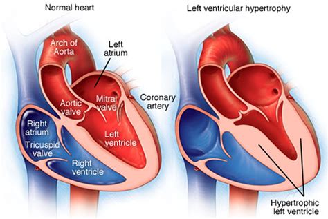
lv enlargement cxr | normal left ventricular enlargement lv enlargement cxr The cardiac silhouette is considered enlarged if the cardiothoracic ratio is . dog / Beds & Furniture / Ramps & Steps. Dog Steps & Dog Ramps. sort by Relevance. Top Paw ® 4-Step Foldable Plastic Steps. $39.99. Extra 20% online only with code SAVE20. Free Same-Day Delivery! Top Paw ® Indoor Foam Steps. $62.99. Extra 20% online only with code SAVE20. Sign In & Enjoy Free Shipping Over $49. Top Paw ® .
0 · normal left ventricular enlargement
1 · left ventricular enlargement ultrasound
2 · left ventricular enlargement radiology
3 · left ventricular enlargement
4 · cardiovascular enlargement
5 · cardiovascular chamber enlargement
6 · cardiomegaly lv enlargement
7 · cardiac enlargement rv
Open the Arduino IDE. Go to the “Tools” menu and select “Manage Libraries.”. In the Library Manager, enter “lvgl” in the search box and press Enter. The Library Manager will display the available LVGL library. Find the “lvgl” library and click the “Install” button on the right. Wait for the installation to complete.
Left ventricularenlargement can be the result of a number of conditions, including: pressure overload. hypertension. aortic stenosis. volume overload. aortic regurgitation. mitral regurgitation. wall abnormalities. left . LV enlargement (LVE) is associated with a wide variety of cardiovascular .
The cardiac silhouette is considered enlarged if the cardiothoracic ratio is .Left Ventricular Enlargement. Assessment of the Size of the left Ventricle (LV) on the Lateral CXR. Lateral examination of a chest x-ray (CXR) shows the normal in the upper row (a,b) and the abnormal and enlarged in the bottom row (c,d).Receiver operating characteristic (ROC) curves for measurements from patients with any . The heart size is considered too large when the CTR is > 50% on a PA chest x .
normal left ventricular enlargement
Left ventricular enlargement tends to round the left heart border as it enlarges laterally and downward. Left atrial enlargement is reflected earliest by enlargement of the appendage on the PA view. More difficult to see is the . This study aimed to evaluate the reliability of the transverse diameter of heart .The left hemidiaphragm should be visible behind the heart. The hemidiaphragm contours do .
versace men eau de parfum
This chapter addresses cardiac enlargement on an AP Chest radiograph. It . Left ventricularenlargement can be the result of a number of conditions, including: pressure overload. hypertension. aortic stenosis. volume overload. aortic regurgitation. mitral regurgitation. wall abnormalities. left ventricular aneurysm. LV enlargement (LVE) is associated with a wide variety of cardiovascular processes, including cardiomyopathy, myocardial infarction, valvular heart disease, PH, and congestive heart failure (25–28). The cardiac silhouette is considered enlarged if the cardiothoracic ratio is greater than 50% on a PA view of the chest 1. See main article: enlargement of the cardiac silhouette for more information.
left ventricular enlargement ultrasound
Left Ventricular Enlargement. Assessment of the Size of the left Ventricle (LV) on the Lateral CXR. Lateral examination of a chest x-ray (CXR) shows the normal in the upper row (a,b) and the abnormal and enlarged in the bottom row (c,d).Receiver operating characteristic (ROC) curves for measurements from patients with any cardiac chamber enlargement, including (A) cardiothoracic ratios (CTR) from posteroanterior chest radiographs (PA-CXR) and (B) anteroposterior chest radiographs (AP-CXR) for both men and women, (C) heart diameters measured from women with (C) PA-CXRs and (D . The heart size is considered too large when the CTR is > 50% on a PA chest x-ray. A CTR of > 50% has a sensitivity of 50% for CHF and a specificity of 75-80%. An increase in left ventricular volume of at least 66% is necessary before it is noticeable on a chest x-ray. On the left a patient with CHF.
Left ventricular enlargement tends to round the left heart border as it enlarges laterally and downward. Left atrial enlargement is reflected earliest by enlargement of the appendage on the PA view. More difficult to see is the posterior bulge on the lateral view just below the carina. This study aimed to evaluate the reliability of the transverse diameter of heart shadow [TDH] by chest X-ray for detecting LV dilatation and dysfunction as compared to Magnetic Resonance Imaging (MRI) performed for different clinical reasons. Materials and Methods:
The left hemidiaphragm should be visible behind the heart. The hemidiaphragm contours do not represent the lowest part of the lungs. Heart size is not assessed by an absolute measurement, but rather is measured in relation to the total thoracic width - the Cardio-Thoracic Ratio (CTR). CTR = Cardiac Width : Thoracic Width. This chapter addresses cardiac enlargement on an AP Chest radiograph. It provides the reader with the basic definition of cardiac enlargement and the contours associated with various chamber enlargements in a question and answer format. Download chapter PDF. Similar content being viewed by others. Keywords. Cardiomegaly. Retrocardiac space.
Left ventricularenlargement can be the result of a number of conditions, including: pressure overload. hypertension. aortic stenosis. volume overload. aortic regurgitation. mitral regurgitation. wall abnormalities. left ventricular aneurysm.
LV enlargement (LVE) is associated with a wide variety of cardiovascular processes, including cardiomyopathy, myocardial infarction, valvular heart disease, PH, and congestive heart failure (25–28).
The cardiac silhouette is considered enlarged if the cardiothoracic ratio is greater than 50% on a PA view of the chest 1. See main article: enlargement of the cardiac silhouette for more information.
Left Ventricular Enlargement. Assessment of the Size of the left Ventricle (LV) on the Lateral CXR. Lateral examination of a chest x-ray (CXR) shows the normal in the upper row (a,b) and the abnormal and enlarged in the bottom row (c,d).Receiver operating characteristic (ROC) curves for measurements from patients with any cardiac chamber enlargement, including (A) cardiothoracic ratios (CTR) from posteroanterior chest radiographs (PA-CXR) and (B) anteroposterior chest radiographs (AP-CXR) for both men and women, (C) heart diameters measured from women with (C) PA-CXRs and (D . The heart size is considered too large when the CTR is > 50% on a PA chest x-ray. A CTR of > 50% has a sensitivity of 50% for CHF and a specificity of 75-80%. An increase in left ventricular volume of at least 66% is necessary before it is noticeable on a chest x-ray. On the left a patient with CHF.Left ventricular enlargement tends to round the left heart border as it enlarges laterally and downward. Left atrial enlargement is reflected earliest by enlargement of the appendage on the PA view. More difficult to see is the posterior bulge on the lateral view just below the carina.
versace mobile price
This study aimed to evaluate the reliability of the transverse diameter of heart shadow [TDH] by chest X-ray for detecting LV dilatation and dysfunction as compared to Magnetic Resonance Imaging (MRI) performed for different clinical reasons. Materials and Methods:The left hemidiaphragm should be visible behind the heart. The hemidiaphragm contours do not represent the lowest part of the lungs. Heart size is not assessed by an absolute measurement, but rather is measured in relation to the total thoracic width - the Cardio-Thoracic Ratio (CTR). CTR = Cardiac Width : Thoracic Width.
left ventricular enlargement radiology
versace medusa stud watch

versace meski niebieski
MEŽTAKA ir garās distances pārgājienu maršruts no Rīgas līdz Tallinai cauri abu valstu mežainākajām teritorijām un trīs nacionālajiem parkiem. Mežtaka ir sadalīta 50 vienas vai divu dienu pārgājiena posmos, kur katrs posms ir ~ 20 km garš. Pārgājienam iespējams izvēlēties jebkuru posmu.
lv enlargement cxr|normal left ventricular enlargement


